What do we see in the retina?
Sonja Moolman - B.Optom (RAU)
Source: https://www.ncbi.nlm.nih.gov/books/NBK11533/
https://www.americaneyeassociates.com/how-does-the-retina-get-its-blood-supply/
Have you ever been to an optometrist who takes a photo of the back of your eye?
Well, most people who see the photos first comment that it looks like Mars or something out of space.
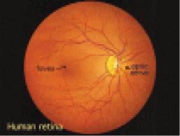
Here is what we as optometrists see when we look at the Retina- back of the eye.
1. OPTIC NERVE:
a. This is the optic nerve that's connected to the brain. A white round like area, where all the blood vessels enter and supply the retina. This nerve is the connection between the eye and the brain, to produce our vision .It consists of more than a million nerve fibers. These nerve fibers carry impulses from the retina to the brain and the brain interprets it as images with colour and contours.
2. BLOOD VESSELS:
a. There are arteries and veins in the retina
Arteries are lighter in colour and supply the retina with oxygenated blood
Arteries are also thinner than the veins
Veins are darker and thicker
b. The central retinal artery enters the eye via the optic nerve. It branches into smaller arteries and supplies all the different parts of the retina, except to the macular area.
b. There is also a layer of blood vessels in the deeper areas of the retina and this supplies the macular area from inside and provides more nutrients to the retina. This inner part is called the Choroid.
3. MACULA (FOVEA)
a. The most important part of your retina. Due to it not having blood vessels, it can have sharper vision. It is the area with the central and fine detail vision.
When taking a photo of the retina, abnormalities can be detected before a serious eye condition can develop such as blindness. It is therefore very important to have your eyes examined at least every 2 year.